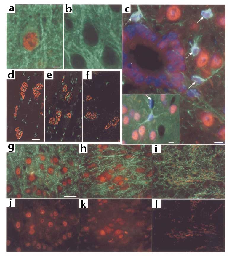
(a) Presence of the Wld protein in neuron nuclei of isocortex and (b) its absence from C57BL/6J control tissue. Red, anti-N70 antibody, which detects Wld and Ube4b. Green, anti-MAP2 antibody markedly outlining neuronal cell bodies. (c) Absence of Wld protein (red) in astrocytes (arrows, main picture). Cytoplasmic staining in ependymal cells (central channel) of WldS thoracic spinal cord could be Ube4b. Inset, absence in astrocytes and endothelial cells (arrow) of WldS isocortex. Green, anti-GFAP. Blue, Hoechst Dye nuclear counterstain. (d-f) Confocal images of triangularis sterni muscle preparations immunostained with anti-N70 antibody (green) and motor endplates counterstained with TRITC alpha-bungarotoxin (red). (d) 4836 homozygote, (e) WldS, (f) C57BL/6J. (g-i) Motor neurons in thoracic spinal cord of both WldS (g) and 4836 (h) expressed the Wld protein (red) in their nuclei, whereas those of C57BL/6J (i) did not. Cytoplasmic signals may be Ube4b or low level Wld protein. Counterstain, neurofilament (green). (j-l) Red channel (Wld protein plus Ube4b) images corresponding to (g-i). Scale bars, 5 mum (a, b), 10 mum (c), 50 mum (d-l).
Font:
Mack TG, Reiner M, Beirowski B, Mi W, Emanuelli M, Wagner D et al. Wallerian degeneration of injured axons and synapses is delayed by a Ube4b/Nmnat chimeric gene. Nat Neurosci. 2001 Dec;4(12):1199-206.